
The Deadlift has always been considered a "Mass Building" exercise. This idea, which was first only supported by the pioneers of bodybuilding, has now begun to be supported by scientific evidence. The deadlift, like other exercises that involve the intervention of compound muscle synergies like the squat and bench press, acts as a noticeable factor of phsyco-physique-metabolic stressogen (or stressor) stimuli, with the stimuli being themselves the input of neuro-umoral stimuli that act as the "Trigger" for the release of hormones of the "Hypotalamus-pituitary-gonad axis" as well as for the "hypothalamus-Somatotrope Cell axis", causing the obvious morphological-functional adaptations always aimed for by the bodybuilder.
These adaptations/modifications act, in part, on one's somatotypic genetic asset, bringing it toward to the aimed muscle growth.
Nowadays the deadlift is performed only by a small group of athletes and experts. This exercise has been long forgotten by all those who have known it and practiced it, and it is now unknown to most beginners. Why are people not using the deadlift? The abandonment of this exercise has its roots in many reasons, with the most common being the deep rooted myth that it is hazardous to the lumbar tract of the spine, or vertebral column. This fear is only justified because of the lack of knowledge in anatomy and kinesiology (which is the study of muscles and their movements) by those who work as pseudo instructors in gyms and fitness centers.
In order to fully comprehend the principles for correct execution and to also learn which muscular benefits are involved, it is required for us to refer to kinesiology. Before we try to comprehend the kinesiology of the deadlift, we must learn a little anatomy and find out which parts of the body are directly involved.
A Brief Overview Of The Anatomy Of The Vertebral Column
The vertebral column is the central pillar of the body and it is made up of a series of short bones that are known as the vertebras (see figure 1 below). There are 32-34 vertebras: 7 cervical, 12 thoracic, 5 lumbar, 5 sacral (fused together forming the Sacrum Bone), and 3-5 coccygeal (fused together forming the Coccyx). The size of the vertebras is based on the function that they serve, with the thickness and the diameter of lumbar vertebras (involved in sustaining the weight of the trunk) is of course greater than those of cervical vertebras that must support only the weight of the head.

Figure 1. Click to enlarge.
The general structure of the vertebra is characterized as following (even though profound differences among vertebras occur in the different tracts of the column):
- It's made up of two major parts: the vertebral body anteriorly and the vertebral arch posteriorly.
- The vertebral arch bears on either side the articular processes which divide the arch into two parts: anteriorly, the pedicles and posteriorly, the laminae;
- The laminae left and right join each other backward to form the spinous process that is attached to the midline posteriorly.
- The vertebral arch is therefore attached to the vertebral body by the pedicles.
- The vertebra also bears transverse processes which are attached to the arch near the articular processes
The vertebra presents a structure formed by a dense bony cortex surrounding a spongy medulla (figure 2). The cortex of the superior and inferior aspects is called the vertebral plateau. On a vertico-frontal plane it's possible to see the spongy centre (figure 2) of the vertebral body with bony trabeculae disposed along the lines of force. These lines are vertical, linking the superior and inferior surfaces, or horizontal, linking the lateral surfaces, or oblique, linking the inferior with the lateral surfaces.
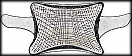
Figure 2.
On a sagittal section (figure 3) it is still possible to notice these trabeculae, but in addition there are two more sheaves of oblique fibres in a fan-like arrangement. The first, (figure 4) arising from the superior surface, fans out at the level of the two pedicles to reach the corresponding superior articular process and the spinous process. The second, (figure 5) arising from the inferior surface, fans out at the level of the two pedicles to reach the corresponding inferior articular processes and the spinous process.
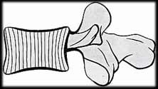
Figure 3.
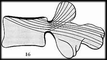
Figure 4.
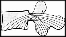
Figure 5.
This criss-crossing of these three trabecular systems constitutes zones of maximum resistance as well as a triangular area of minimum resistance, made up entirely of vertical trabeculae (figure 6).
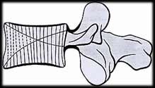
Figure 6.
Having seen the structure and the lines of force of the vertebra, this explains why the anterior part of the vertebral body is less resistant to the axial compression forces than it's posterior part. In fact an axial compression force of 600 kg (1320 lbs) will crush the anterior part of the vertebral body leading to a compression fracture. Instead an axial compression force of 800 kg (1760 lbs) is required to crush the whole vertebra and make the posterior part give way (figure 7).
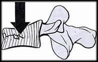
Figure 7.
The joint that is located between two vertebrae is an anphyarthrosys. It is made up of two vertebral plateaus that are connected by the intervertebral disc.
The intevertebral disc consists of a central part and a peripheral part (figure 8).

Figure 8. Click to enlarge.
The central part, the nucleus pulposus, a gelatinous substance containing 88% water.
The peripheral part, the annulus fibrosus, is formed by concentric fibres which appear to cross one another obliquely in space. The fibres are vertical peripherally and become more oblique towards the centre.
Thus the nucleus is enclosed within an inextensible casing made up by the vertebral plateaus and the annulus.
Now that you understand a little about the basic anatomy of the spine, let's see how to perform this exercise correctly and find out what the major mistakes are.
CORRECT EXECUTION
Starting position and beginning of the movement (figure 9):
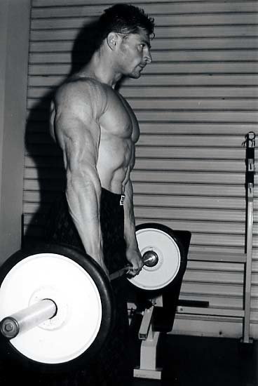
Figure 9: Beginning. Click to enlarge.
Starting with the torso erect and your legs opened as wide as your shoulder width, grasp the barbell with both hands in pronation (overhand grip), or one prone and the other one supine (underhand grip). Flex your trunk while keeping the vertebral column aligned on the same axis and bring the barbell slightly downward below your knee level until the back is parallel to the ground and without the presence of any convex curve posteriorly at the thoracic-lumbar as well as at the lumbar-sacral level.
During the flexion of the trunk (figure 10) it is required that you pay attention to three important factors:
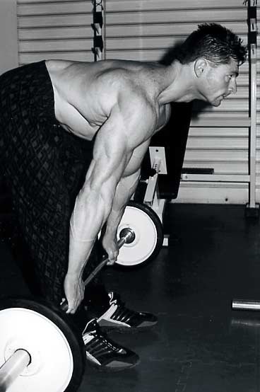
Figure 10: End. Click to enlarge.
- Keep all the vertebral segments of the column aligned. In order to elicit this condition it is useful to keep the face looking ahead, instead of looking downward, keeping the head in hyperextension while flexing the trunk.
- Keep the barbell as close as possible to your legs during the downward phase of the movement.
- Inhale deeply during the downward-eccentric phase (while flexing the trunk) and keep the inhaled air in (without exhaling it) until half of the subsequent upward-concentric phase has been reached with the contemporaneous abdominal contraction in order to elicit the "Valsalva Maneuvor".
Final phase and return to the starting position:
The returning phase consists of the opposite of flexion. It is required that you pay attention to the exhalation; exhaling progressively (start exhaling from half of the concentric-upward phase) until the end of the complete vertebral column extension, or in other words, when you are standing straight up.
INCORRECT EXECUTION
The main mistakes in the incorrect execution of this exercise are:
- Flexion of the vertebral bodies (one over each other) instead of keeping them aligned during the flexion of the trunk (figure 11)
- Inhalation and exhalation performed inversely or premature (early) expiration during the concentric-upward phase
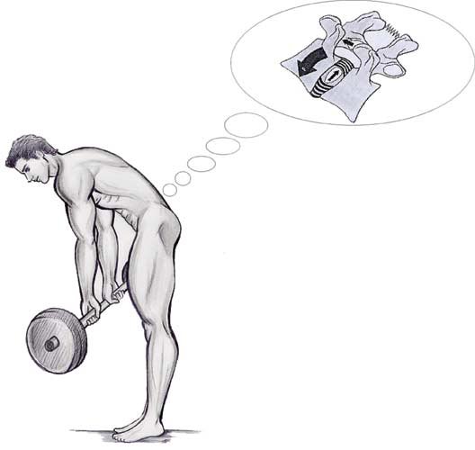
Figure 11. Click to enlarge.
Now that we have mentioned the principal motivations behind the correct and incorrect execution of this exercise, it's even more important to give an explanation from a kinesiological point of view.
1. The importance of vertebras alignment and of vertebral column segments during the flexion of the trunk.
The nucleus has a so called "Preloaded state", and therefore even without any axial compression there exists a base tension of the annulus fibrosus fibres induced by this nucleus preloaded state. When an asymmetrical load is applied axially to a disc the upper vertebral plateau tilts toward the overloaded side (figure 12), and so the fibre AB' will be stretched to AB. This asymmetrical axial compression also acts indirectly over the nucleus, which has a preloaded state and so it moves toward that side (direction of the arrow) where the intervertebral space opens and thus the nucleus will bring back the fibre to the position AB', thereby righting the vertebral plateau and restoring it to it's original position. This is the so called "mechanism of self-stabilization" which is highly dependent by the cooperation of the couple nucleus-annulus.
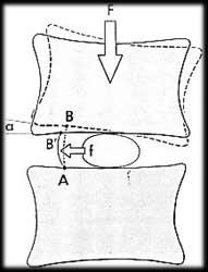
Figure 12.
During extension movements (figure 13) the body of the upper vertebra tilts and moves posteriorly, so that consequently the thickness of the disc is reduced posteriorly and expanded anteriorly, and the nucleus is pushed anteriorly, and then leans towards the anterior fibres of the annulus augmenting their tension state and therefore consequently bringing it back to it's initial position.
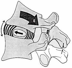
Figure 13. Click to enlarge.
During flexion movements (figure 14) the body of the upper vertebra tilts and slides anteriorly reducing the thickness of the intervertebral disc anteriorly and increasing it posteriorly; the nucleus is pushed backward against the posterior fibres of the annulus augmenting their tension state.
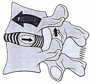
Figure 14. Click to enlarge.
Therefore if during the flexion of the trunk the vertebras are not kept aligned and therefore they flex one over each other, during the descendent-eccentric phase of the Deadlift, the thickness of the intervertebral discs get reduced anteriorly and causes the posterior intervertebral space to open and consequently the migration of the nucleus backward (figure 15 A).
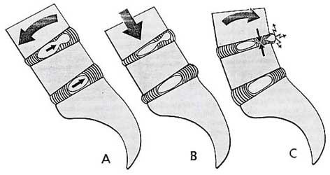
Figure 15. Click to enlarge.
Immediately following the descendent-eccentric phase of the Deadlift, there's the lifting effort and at the beginning of this phase the axial compression crushes the whole disc and violently drives the nuclear substance backward until it reaches the deep surface of the posterior longitudinal ligament (figure 15 B).
During the extension of the vertebral column (in the upward-concentric phase of the Deadlift that allows it to get back to the starting position) and the extension of the vertebras one over each-other cause the closing of the intervertebral space posteriorly and it's opening anteriorly with the obvious migration (for the reasons explained earlier) of the nucleus anteriorly.
When you are back to the starting position, at the point of complete extension of the vertebral column, the closing of the posterior intervertebral space could promote (in the case of presence of microtraumas of the annulus fibrosus) the non complete migration of the nucleus anteriorly. This causes a part of the nucleus to remain trapped under the posterior longitudinal ligament and therefore causes the formation of a herniating mass because of the cutting effect induced by the violent closing of the posterior intervertebral space (figure15 C).
It's obvious that in order to prevent traumas to the vertebral column, when performing this exercise one must avoid emphasizing the sliding of the vertebras (that are adjacent) one over each other during the flexion of the trunk.
2. The Importance Of Keeping The Barbell Close To The Legs
During forward flexion the weight of the trunk along with the weight of the head apply to the partial center of gravity (P) placed just anteriorly to the 10th thoracic vertebrae. This weight is applied at the tip of the long arm of a lever with its fulcrum at the level of the nucleus pulposus of L5-S1.
To counterbalance this force the paravertebral muscles (S1) that are acting on the short arm of the lever, which is 7 to 8 times shorter than the long arm, must develop a force 7 to 8 times greater than P1 (figure 16). This means the force acting on the lumbosacral disc is equal to S1+P1.
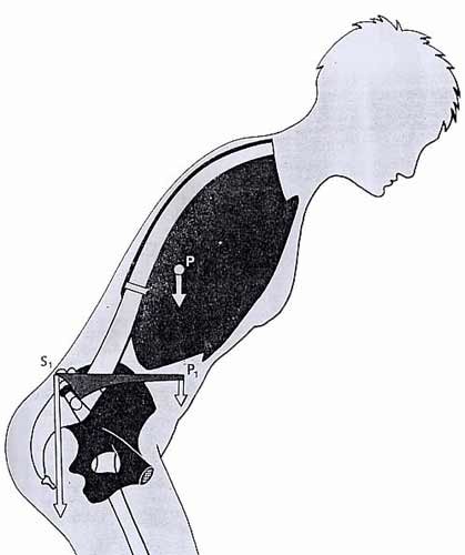
Figure 15. Click to enlarge.
This force will be even greater as much as you increase the flexion of the trunk forward since flexion increases the distance of the tip of the arm of the lever P1 from its base S1. The force acting on the lumbosacral disc increases even more when you carry weights in your hands with your arms outstretched anteriorly.
At this point it's obvious that if you don't want to avoid excessive flexion of the trunk forward (because of it's great stimulation of the Gluteus Maximus muscle and the back of the thigh muscles during the upward-concentric phase) it's important to exclude any excessive overload caused by a useless frontward position of the arms.
3. Importance Of Respiration And Abdominal Contraction For Injury Prevention
The correct respiration while performing this exercise contributes not only to the fixation of the chest cavity which helps you to better sustain the weight lifted, but it's also the main factor involved in keeping your vertebral column safe and helping you to avoid fracturing the vertebral bodies in extreme cases.
Let's take a detailed look on how this statement is justified.
To lift a weight of 10 kg, with your knees flexed and your trunk held vertically, the force S1 exerted by the paravertebral muscles equals 141 kg. Lifting the same weight with extended knees and trunk flexed forward, requires a force of 256 kg. If the same weight is carried with the arms outstretched anteriorly, S1 equals 363 kg. According to some authors, the force acting on the nucleus ranges from 282 to 726 kg or even up to 1200 kg with the latter value clearly exceeding the force required to smash the vertebral bodies (800 kg in the posterior part, the more resistant part; 600 kg in the anterior part, the less resistant part).
The resistance of vertebral bodies to such axial compression is justified by two factors that clarify this contradiction:
- The weight on the intervertebral disc is well distributed so that the nucleus bears 75% of the force and the annulus only 25%
- The second factor, most likely the most important, is that one that is influenced by our RESPIRATION, the inhalation during the descendent-eccentric phase and the following exhalation during the following upward-concentric phase constitute the correct respiratory procedure to be adopted while performing every kind of exercise (except for a few).
After the descendent-eccentric phase in which you inhale, it is extremely important to keep the inhaled air in (in other words,remaining in an apnoea state) for the execution of the first phase of the upward-concentric phase. After this, during the rest of the upward-concentric phase, the exhalation must be performed simultaneously at the closing of the glottis and all the abdominal orifices (Valsalva Manoeuvre) in order to increase the pressure within the thoraco-abdominal cavity followed by the abdominal contraction.
The pressure then rises considerably within the cavity and transforms it into a rigid beam lying anterior to the vertebral column and transmitting the forces exerted on the bony pelvis and the perineum.
The functional utility of this rigid beam reverberates positively on reducing the axial compression forces acting on the intervertebral discs; in detail this compression gets reduced by 50% at the level of the T12-L1 disc, and 30% at the level of the L5-S1 disc, consequently the tension of paravertebral muscles gets reduced by 55% also.
If at one side the Valsalva Manoeuvre allows you to preserve the vertebral column from the risk of undesired injuries, but on the other side this Manoeuvre is not without physiological consequences. In fact it determines cardio vascular modifications. The increase of pressure created by this manoeuvre is accompanied with an increase of the intrathoracic pressure which compress the major veins (Superior Vena Cava and Inferior Vena Cava) that lead the blood back to the heart.
A reduction in venous return determines a lesser filling of the Right Ventricle, that reflects itself in a consequently lesser filling of the Left Ventricle. Therefore the Left Ventricle, exerting a low arterial pressure, is not able to satisfy an optimal perfusion of the organs, and among them the BRAIN. This condition produces dizziness, "spots before the eyes", and even fainting with straining-type exercises. Normal blood flow is re-established (with perhaps even an "overshoot" in flow) once the glottis is opened and intrathoracic pressure is released.
This is pointed out in order to remind you that an optimal hypertrophic stimulus, without compromising your conscience state, is satisfied with a range of 8 to 12 reps.
Another useful tip concerns the correct positioning of your feet in order to promote and not interfere with the automatic physiological axial rotation of the knees. When the knee is extended the foot is laterally rotated and when it's flexed the foot is medially rotated. Therefore, while performing the Deadlift (especially the form with knees extended), it is important to place the feet so that the toes face slightly outwards-laterally. This is necessary for helping the lateral rotation of the leg that occurs when the knee is fully extended.
Muscle Interventions
The main muscles that are directly involved while performing the Deadlift are:
 LEG POSTERIOR MUSCLES (CALVES):
- Gastrocnemius muscle
- Gastrocnemius muscle (lateral head)
- Gastrocnemius muscle (medial head)
- Soleus muscle
- Popliteus muscle
THIGH POSTERIOR MUSCLES (HAMSTRINGS):
- Bicep femoris
- Long head of bicep femoris
- Short head of bicep femoris
- Semitendinosus muscle
- Semimembranosus muscle
GLUTEUS (BUTT):
- Gluteus maximum muscle
- Gluteus medius muscle
- Gluteus minimum muscle
LUMBAR MUSCLES (LOWER BACK):
- Sacrus-spinosus muscle
- Spinosus muscle
- Transversi-spinosus muscle
- Interspinalis lumborum muscles
The order in which the muscle groups have been listed reflects either the order they are disposed anatomically from downward to upward and the order of their recruitment from the beginning of the concentric-upward phase of the exercise.
Why The Deadlift Is One Of The Best Exercises
Now, let's see why the deadlift can be considered one of the best exercises for your Gluteus muscles and hamstrings.
Gluteus maximus muscle: This muscle acts as a hip extensor either when it has its fixed point at the proximal origin and when it has its fixed point at the distal insertion.
Therefore its recruitment occurs either while bringing the femur backward (i.e. while performing the Gluteus machine) and in all those movements that involve the straightening of the trunk after being flexed downward (i.e. deadlift).
The Gluteus maximus muscle is better stimulated during the straightening of the trunk than the extension of the lower limb backward. This is because when the hip goes through a higher degree of flexion (in other words, tilted forward) the fibres of Gluteus maximus muscle lengthen to100%, and the more the lengthening the more powerful the contraction during the following phase of hip extension; furthermore the power of contraction is even more efficient the greater the degree of knee extension (i.e. with the trunk bent forward and the hands touching the feet, as in the position at the end of the eccentric-descendent phase of the deadlift).
Hamstrings: These three muscles (biceps femoris, semitendinosus, semimembranosus) are knee flexors when they have their fixed point at the proximal origin (i.e.: while performing the leg curl exercise). They also act as hip extensors when they have their fixed point at the distal insertion (i.e.: deadlift).
During the flexion of the hip (in the case of the deadlift during the eccentric-descendent phase) the distance between origins and insertions increases progressively. This means that the more the hip is flexed, the greater the degree of relative shortening of the hamstrings and consequently the more stretched they become. All this turns in a greater power of contraction during the following hip extension phase (during the straightening of the trunk like during the concentric-upward phase of the deadlift).
Straight Legs Or Bent Legs?
Deadlift with straight, extended knees: During the eccentric-descendent phase the degree of forward flexion of the trunk is greater, and therefore the partial center of gravity, fixes itself on a longer arm of a lever that involve a greater stress of paravertebral muscles that act on a shorter arm of a lever. Under a muscular point of view, during the concentric upward-phase, the short head of biceps femoris don't take part in the hip extension during the straightening of the trunk, because it's a monoarticular muscle.
Deadlift with bent (or half bent) knees: During the eccentric-descendent phase the degree of forward flexion of the trunk is lesser than seen with extended knees because of the bending of the knees; and the more bent the knees are, the lesser the degree of forward flexion of the trunk and consequently lesser the stress beared by the paravertebral muscles.
Under a muscular point of view, the lesser flexion of the trunk forward will determine a lesser "relative shortening" of the Gluteus maximus muscle and Hamstrings and consequently a lesser stimulation of them during the following concentric-upward phase (if compared to the Deadlift performed with extended knees). If during the straightening of the trunk the knees are kept bent, it will cause the muscular intervention of the calves, popliteus muscle and short head of bicep femoris that help in maintaining the bent position of the knee for the whole range of movement (because they are knee flexors).
Summary And Conclusion
So to sum up the main points:
- The deadlift is a mass building exercise which causes "muscle building" hormones to be released by your body.
- It is important to understand the kinesiology of the exercise, as described in detail above.
- Correct form is very important. The most important things to watch for are keeping your spine straight throughout the entire exercise, keeping the barbell close to your legs at all times, and inhaling during the downward phase, and exhaling once you are halfway back up to the standing position.
- The deadlift works the calves, glutes, hamstrings, and lower back.
I hope to have made clear the necessary fundamentals for the correct execution of the deadlift so that this exercise can be re-evaluated and no longer forgotten. I wish great growth to everybody